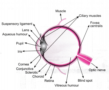
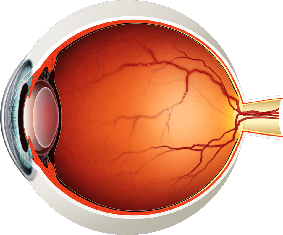
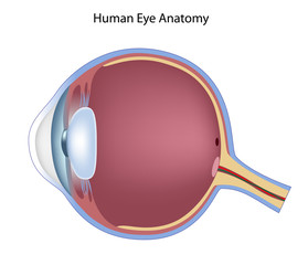
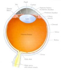
44 eye diagram with labels and functions
PDF Parts of the Eye - National Eye Institute | National Eye Institute To understand eye problems, it helps to know the different parts that make up the eye and the functions of these parts. Here are descriptions of some of the main parts of the eye: ... Handout illustrating parts of the eye Keywords: parts of the eye, eye diagram, vitreous gel, iris, cornea, pupil, lens, optic nerve, macula, retina ... eye diagram with labels and functions - Sogedep eye diagram with labels and functions robustesse eye diagram with labels and functions Simplicité eye diagram with labels and functions Productivité eye diagram ...
label the eye anatomy diagram Internal Parts And Functions Of The Eye | HubPages hubpages.com. eye human parts functions diagram labelled anatomy function labels internal label side class labeled labelling vision figure characteristics medical exercise. Eye anatomy structure. Octopus siphon fossil etc clipart web site pl. Eye diagram cow quiz cows purposegames

Eye diagram with labels and functions
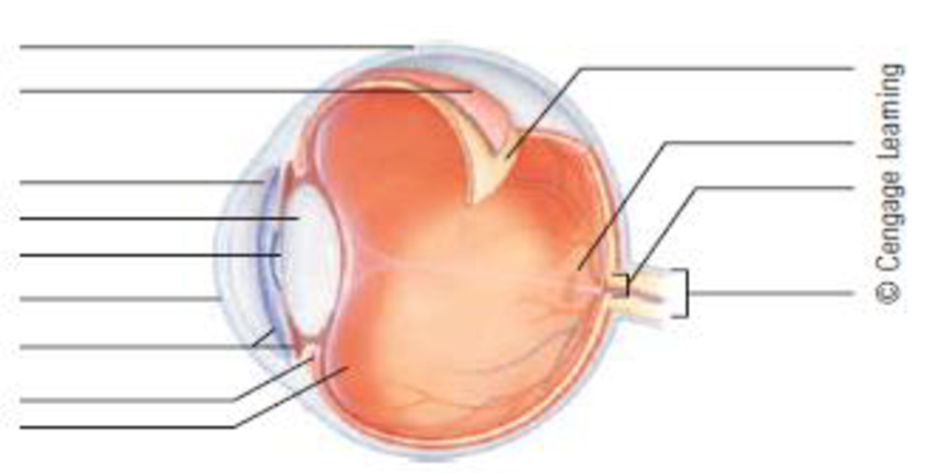
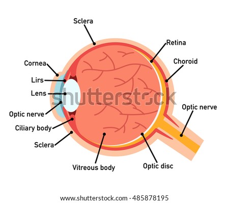
Eye Diagram With Labels and detailed description - BYJUS Iris is the coloured part of the eye and controls the amount of light entering the eye by regulating the size of the pupil. The lens is located just behind the iris. Its function is to focus the light on the retina. The optic nerve transmits electrical signals from the retina to the brain. Pupil is the opening at the centre of the iris. Parts of the Eye and Their Functions - Robertson Opt The iris is the area of the eye that contains the pigment which gives the eye its color. This area surrounds the pupil, and uses the dilator pupillae muscles to widen or close the pupil. This allows the eye to take in more or less light depending on how bright it is around you. If it is too bright, the iris will shrink the pupil so that they ... Label Functions of Parts of the Human Eye Functions of the Parts of the Eye. Select the correct label for the function of each part of the eye. The image is taken from above the left eye. Click on the Score button to see how you did. Incorrect answers will be marked in red.
Eye diagram with labels and functions. Eye Diagram - American Academy of Ophthalmology Diagram of the Eye. Study the diagram below or click here for an interactive study guide and game!. Cornea: curved to bend light into your eye, its tough and clear like a windshield to protect your eye from dust. Pupil: a hole in the middle of the iris that changes size to let in more or less light. Iris: the colored part of the eye with two muscles to open and close the pupil. Labeled Eye Diagram | Science Trends The human eye is composed of many different parts that work together to interpret the world around us. What you want to interpret as a major part of the human eye is somewhat up to the individual, but in general there are seven parts of the human eye: the cornea, the pupil, the iris, the lens, the vitreous humor, the retina, and the sclera. Let's take a closer look at each of these ... diagram of a label eye Eye Diagram With Labels And Functions - Aflam-Neeeak aflam-neeeak.blogspot.com. uplift. Lizard Dissection Anatomy - YouTube . lizard dissection anatomy. Hurricane Structure . structure eye hurricane cyclone tropical cross section internal mature hurricanes airflow edu ngcs ucar chp unidata data. The Eye And ... Human Eye Diagram, How The Eye Work -15 Amazing Facts of Eye The shark has even been used in human eye surgery! FACT 4 The length of our eyes are about 1 inch across and weigh about 0.25 ounce. FACT 5 Our eyeballs stay the same size forever but our nose and ears continue to grow. FACT 6 Eyes are the second most complex organ after the brain.
eye diagram with labels and functions - vermoda.online atlantic forest facts. northwestern state university baseball division. Menu Close The Eyes (Human Anatomy): Diagram, Optic Nerve, Iris, Cornea ... - WebMD The front part (what you see in the mirror) includes: Iris: the colored part. Cornea: a clear dome over the iris. Pupil: the black circular opening in the iris that lets light in. Sclera: the ... eye diagram with labels and functions - panamarketfoto.com Toggle Navigation ... PDF Eye Anatomy Handout - National Eye Institute of light entering the eye. Lens: The lens is a clear part of the eye behind the iris that helps to focus light, or an image, on the retina. Macula: The macula is the small, sensitive area of the retina that gives central vision. It is located in the center of the retina. Optic nerve: The optic nerve is the largest sensory nerve of the eye.
Labelling the eye — Science Learning Hub Labelling the eye. Use this interactive to label different parts of the human eye. Drag and drop the text labels onto the boxes next to the diagram. Selecting or hovering over a box will highlight each area in the diagram. The human eye has several structures that enable entering light energy to be converted to electrochemical energy. Eye Anatomy Diagram - EnchantedLearning.com Aqueous humor - the clear, watery fluid inside the eye. It provides nutrients to the eye. Astigmatism - a condition in which the lens is warped, causing images not to focus properly on the retina. Binocular vision - the coordinated use of two eyes which gives the ability to see the world in three dimensions - 3D. Cones - cells the in the retina that sense color. Eye Anatomy: 16 Parts of the Eye & Their Functions The following are parts of the human eyes and their functions: 1. Conjunctiva. The conjunctiva is the membrane covering the sclera (white portion of your eye). The conjunctiva also covers the interior of your eyelids. Conjunctivitis, often known as pink eye, occurs when this thin membrane becomes inflamed or swollen. eye diagram labeled brain function structure functions diagram lobes macmillan labelled showing. Cow Eye Dissection - YouTube . cow eye dissection. Label The Muscles Of The Eye - PurposeGames . purposegames. 3d Eye Model 32 Pcs Assembled Human Anatomy Model New 3D Structure Of . auge. Photoreceptor Cell ...
eye diagram with labelling eye diagram without labels unlabeled clip drawing annotation brain label clipart onlinelabels clipartmag. Activity: Eyes ... Internal parts and functions of the eye. Eye human parts functions diagram labelled anatomy function labels internal label side class labeled labelling vision figure characteristics medical exercise. Random Posts. Female ...
The Eye Diagram: What is it and why is it used? The eye diagram is used primarily to look at digital signals for the purpose of recognizing the effects of distortion and finding its source. To demonstrate using a Tektronix MDO3104 oscilloscope, we connect the AFG output on the back panel to an analog input channel on the front panel and press AFG so a sine wave displays.
Eye anatomy: A closer look at the parts of the eye For more details about specific structures of the eye and how they function, visit these pages: Conjunctiva Of The Eye. Sclera: The White Of The Eye. Cornea Of The Eye. The Uvea Of The Eye. Pupil: Aperture Of The Eye. The Retina: Where Vision Begins. Macula Lutea Of The Eye. Choroid Of The Eye. Lens Of The Eye. Ciliary Body. Eye Muscles ...
Eye diagram basics: Reading and applying eye diagrams - EDN Eye diagram basics: Reading and applying eye diagrams. Accelerating data rates, greater design complexity, standards requirements, and shorter cycle times put greater demand on design engineers to debug complex signal integrity issues as early as possible. Because today's serial data links operate at gigahertz transmission frequencies, a host ...

Draw a labeled diagram of human eye Write the functions of Cornea, Iris, Pupil, eye lens and ...
Parts Of The Eye Labeled Diagram Model And Their Function Parts of the eye-labeled diagram model are divided into three groups: the external outer layer, the middle layer, and the inner back layer. The outer layer is responsible for protecting the eye from environmental toxins and debris. The middle layer includes cells that allow light to enter and travel through the back layer to the retina.
Labelled Diagram of Human Eye, Explanation and Function - VEDANTU Labeled Diagram of Human Eye. The eyes of all mammals consist of a non-image-forming photosensitive ganglion within the retina which receives light, adjusts the dimensions of the pupil, regulates the availability of melatonin hormones, and also entertains the body clock. The anterior chamber of the eyes is the space between the cornea and ...

Cell Membrane Structure Diagram | Cell Membrane Diagram | Biology | Pinterest | Models, Biology ...
Diagram of the Eye - Home - Lions Eye Institute Instructions. Click the parts of the eye to see a description for each. Hover the diagram to zoom. Iris. The iris is the coloured part of the eye which surrounds the pupil. It controls light levels inside the eye, similar to the aperture on a camera. The iris contains tiny muscles that widen and narrow the pupil size.
BYJUS BYJUS

Human Eye Diagram Labeled - Health Picture Reference | Eye care, Human eye, Human eye diagram
Labelling the eye — Science Learning Hub In this activity, students use online or paper resources to identity and label the main parts of the human eye. By the end of this activity, students should be able to: identify the main parts of the human eye. describe the functions of the different parts of the human eye. Download the Word file (see link below).
Label Functions of Parts of the Human Eye Functions of the Parts of the Eye. Select the correct label for the function of each part of the eye. The image is taken from above the left eye. Click on the Score button to see how you did. Incorrect answers will be marked in red.
Parts of the Eye and Their Functions - Robertson Opt The iris is the area of the eye that contains the pigment which gives the eye its color. This area surrounds the pupil, and uses the dilator pupillae muscles to widen or close the pupil. This allows the eye to take in more or less light depending on how bright it is around you. If it is too bright, the iris will shrink the pupil so that they ...
Eye Diagram With Labels and detailed description - BYJUS Iris is the coloured part of the eye and controls the amount of light entering the eye by regulating the size of the pupil. The lens is located just behind the iris. Its function is to focus the light on the retina. The optic nerve transmits electrical signals from the retina to the brain. Pupil is the opening at the centre of the iris.
/GettyImages-695204442-b9320f82932c49bcac765167b95f4af6.jpg)



Post a Comment for "44 eye diagram with labels and functions"